Abstract
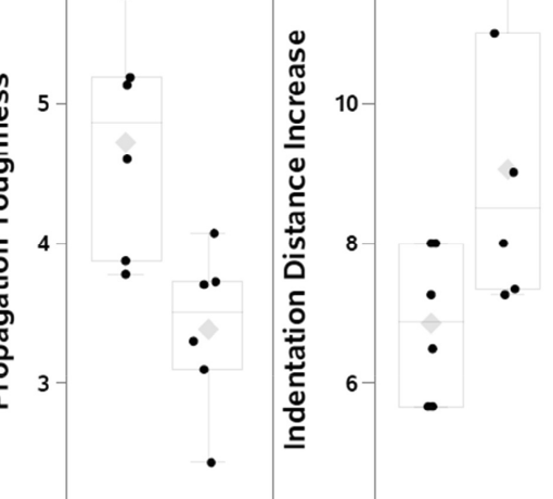
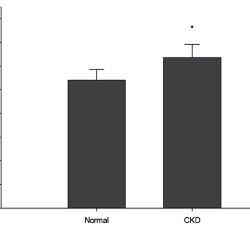
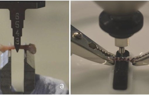
Osteogenesis imperfecta is a congenital disease commonly characterized by brittle bones and caused by mutations in the genes encoding Type I collagen, the single most abundant protein produced by the body. The oim model has a natural collagen mutation, converting its heterotrimeric structure (two α1 and one α2 chains) into α1 homotrimers. This mutation in collagen may impact formation of the mineral, creating a brittle bone phenotype in animals. Femurs from male wild type (WT) and homozygous (oim/oim) mice, all at 12 weeks of age, were assessed using assays at multiple length scales with minimal sample processing to ensure a near-physiological state. Atomic force microscopy (AFM) demonstrated detectable differences in the organization of collagen at the nanoscale that may partially contribute to alterations in material and structural behavior obtained through mechanical testing and reference point indentation (RPI). Changes in geometric and chemical structure obtained from µ-Computed Tomography and Raman spectroscopy indicate a smaller bone with reduced trabecular architecture and altered chemical composition. Decreased tissue material properties in oim/oim mice are likely driven by changes in collagen fibril structure, decreasing space available for mineral nucleation and growth, as supported by a reduction in mineral crystallinity. Multi-scale analyses of this nature offer much in assessing how molecular changes compound to create a degraded, brittle bone phenotype.
https://www.ncbi.nlm.nih.gov/pubmed/25158170
Connect Tissue Res. 2014 Aug;55 Suppl 1:4-8. doi: 10.3109/03008207.2014.923860.


